
- The one and a half syndrome is characterized by a conjugate horizontal gaze palsy in one direction and an impairment of the abduction of the eyes in the other. (T/F?)
- Horizontal gaze palsy is seen on looking away from the side of the lesion. (T/F?)
- The ipsilateral eye has no horizontal movement. (T/F?)
- The most common cause of the one-and-a-half syndrome in the young people is Miller-Fisher syndrome. (T/F?)
- The most common cause of the one-and-a-half syndrome in older people is multiple sclerosis. (T/F?)
- When the lesion that causes a damage to PPRF or abducens nucleus and medial longitudinal fasciculus, also affects the facial nerve, we can talk about an eight-and-a-half syndrome. (T/F?)
80. Corticospinal tract (Gray764)
79. The blood-brain barrier (BBB)
- The blood–brain barrier (BBB) is a nonpermeable membrane that separates the circulating blood from the brain and extracellular fluid in the central nervous system (CNS). (T/F?)
- The blood–cerebrospinal fluid barrier (BCSFB) is a barrier that separates blood from the cerebrospinal fluid (CSF). (T/F?)
- Capillary endothelial cells in the brain are linked by tight junctions which normally do not exist in the rest of the circulation. (T/F?)
- Area postrema and amygdala are parts of the brain that lack normal blood-brain barrier. (T/F?)
- When the meninges are inflamed the blood-brain barrier can be disrupted, which can increase the penetration of the antibiotics into the brain tissue. (T/F?)
- Disruption of the blood-brain barrier is characteristic for myasthenia gravis. (T/F?)
78. Blood supply of the central nervous system (I)
- Parts of the primary motor cortex that control movement of the legs are supplied by branches of the anterior cerebral artery. (T/F?)
- The recurrent artery of Heubner is a branch of the anterior cerebral artery. (T/F?)
- The anterior choroidal artery originates from the anterior cerebral artery. (T/F?)
- The Vidian artery is the artery of the pterygoid canal. (T/F?)
- The Broca’s area is supplied by the branches of the anterior cerebral artery. (T/F?)
- The primary visual cortex is supplied by branches of the posterior cerebral artery. (T/F?)
77. Circle of Willis (Gray-519)

76. Circle of Willis
- Terminal branches of the internal carotid artery are the anterior cerebral artery and the median cerebral artery. (T/F?)
- The anterior communicating artery connects the two anterior cerebral arteries. (T/F?)
- Arterial branches from the anterior communicating artery supply optic chiasm with arterial blood. (T/F?)
- The posterior communicating artery connects the middle cerebral artery and the posterior cerebral artery. (T/F?)
- The pontine arteries arise from the posterior communicating arteries. (T/F?)
- The posterior cerebral artery arises from the basilar artery. (T/F?)
75. Branches of the internal carotid artery
- Anterior choroidal artery. (T/F?)
- Anterior hypophyseal artery. (T/F?)
- Posterior hypophyseal artery. (T/F?)
- Superior hypophyseal artery. (T/F?)
- Anterior communicating artery. (T/F?)
- Posterior communicating artery. (T/F?)
74. Entry and exit points of cranial nerves, arteries and veins (I)
- Supraorbital artery: Supraorbital foramen. (T/F?)
- Ophtalmic nerve: optic canal. (T/F?)
- Mandibular nerve: foramen ovale. (T/F?)
- Middle meningeal artery: foramen spinosum. (T/F?)
- Accessory nerve: foramen ovale. (T/F?)
- Hypoglossal nerve: jugular foramen. (T/F?)
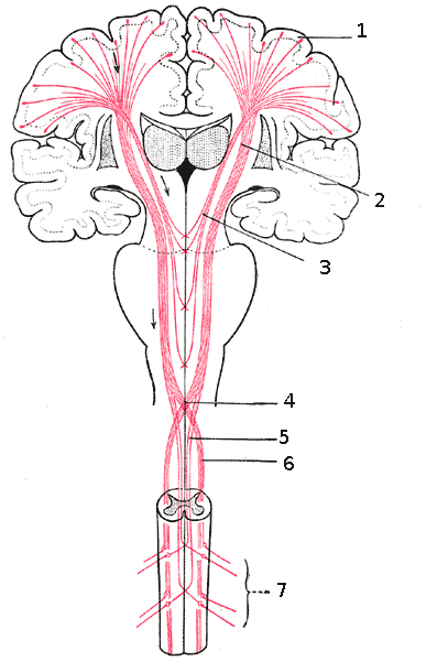
73. Descending neural pathways
- The posterior funiculus contains two descending tracts: the interfascicular fasciculus and the septomarginal fasciculus. /T/F?)
- The lateral corticospinal tract is organized somatotopically, cervical segments are located laterally and sacral segments medialy. (T/F?)
- The rubrospinal tract is situated in the lateral funiculus of the spinal cord. (T/F?)
- The lateral vestibulospinal tract runs the entire length of the spinal cord. (T/F?)
- The medial vestibulospinal tract runs down only to the cervical segments of the spinal cord. (T/F?)
- Spinospinal tracts are collections of fibers that connect various levels of the spinal cord and they are present in all spinal funiculi (posterior, lateral and anterior). (T/F?)
72. Sensory pathways (II)
- The thalamus contains the second order neurons of the sensory pathways. (T/F?)
- The cuneate fasciculus (fasciculus cuneatus) is located in the posterior white column of the spinal cord and carries information from the lower limbs. (T/F?)
- The posterior and anterior spinocerebellar tracts and the lateral spinothalamic tract are located in the lateral funiculus of the spinal cord. (T/F?)
- The spinocerebellar tracts carry unconscious proprioception from the whole body to the cerebellum. (T/F?)
- The anterior spinothalamic tract and the spino-olivary tract are located in the anterior funiculus. (T/F?)
- The spino-olivary tract carries proprioception information from muscles and tendons to the olive. (T/F?)